Figure 2. Immunohistochemical staining for
stem cell factor (SCF). Vascular endothelial cells and stromal cells
expressed strong immunoreactivity for SCF in a membrane from a patient
with active proliferative diabetic retinopathy (PDR). A: Low
power, original magnification 40×. B: high-power, original
magnification 100×. SCF immunoreactivity is absent in a membrane from a
patient with inactive PDR. Note that the membrane is composed mostly of
fibrous tissue (C: original magnification 40×).
Immunohistochemical staining for granulocyte colony-stimulating factor
(G-CSF). G-CSF immunoreactivity was observed in vascular endothelial
cells (D: original magnification 100×). Immunohistochemical
staining for endothelial nitric oxide synthase (eNOS). Immunoreactivity
for eNOS was observed in vascular endothelial cells (E: original
magnification 100×) and stromal cells (F: Low-power, original
magnification 40× and G: high-power, original magnification
100×).
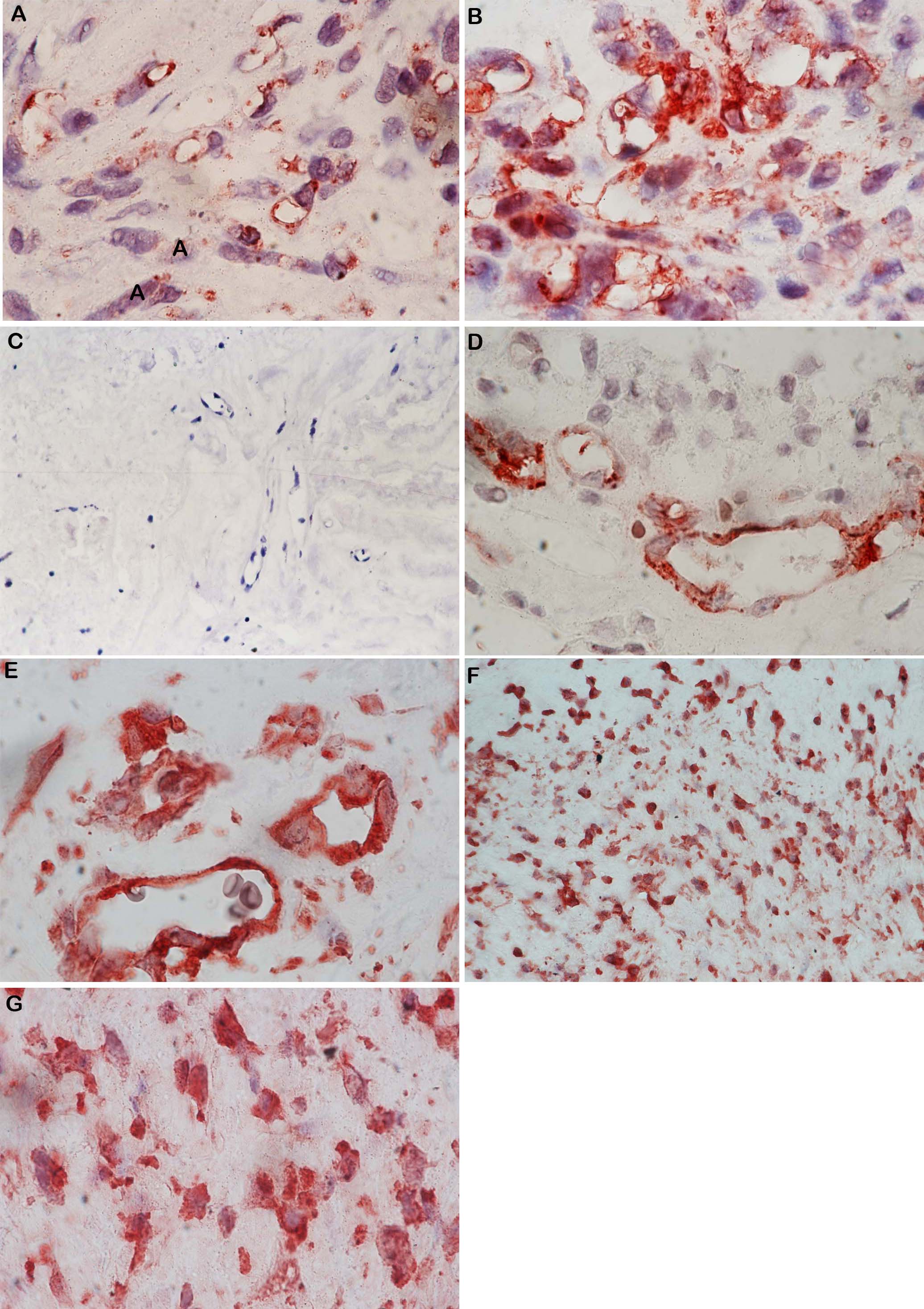
 Figure 2 of Abu El-Asrar, Mol Vis 2010; 16:1098-1107.